|
|
|
|
Generation of the Heartbeat
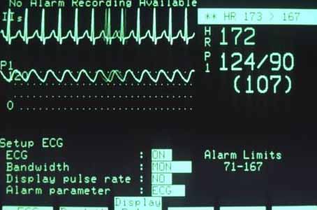
Electrocardiograph An electrocardiograph (ECG or EKG) records the electrical activity of the heart. Preceding each contraction of the heart muscle is an electrical impulse generated in the sinoatrial node; the waves displayed in an ECG trace the path of that impulse as it spreads through the heart. Irregularities in an ECG reflect disorders in the muscle, blood supply, or neural control of the heart.Photo Researchers, Inc./Hank Morgan/Science Source
Unlike most muscles, which rely on nerve impulses to cause them to contract, heart muscle can contract of its own accord. Certain heart muscle cells have the ability to contract spontaneously, and these cells generate electrical signals that spread to the rest of the heart and cause it to contract with a regular, steady beat.
The heartbeat begins with a small group of specialized muscle cells located in the upper right-hand corner of the right atrium. This area is known as the sinoatrial (SA) node. Cells in the SA node generate their electrical signals more frequently than cells elsewhere in the heart, so the electrical signals generated by the SA node synchronize the electrical signals traveling to the rest of the heart. For this reason, the SA node is also known as the heart’s pacemaker.
Impulses generated by the SA node spread rapidly throughout the atria, so that all the muscle cells of the atria contract virtually in unison. Electrical impulses cannot be conducted through the partition between the atria and ventricles, which is primarily made of fibrous connective tissue rather than muscle cells. The impulses from the SA node are carried across this connective tissue partition by a small bridge of muscle called the atrioventricular conduction system. The first part of this system is a group of cells at the lower margin of the right atrium, known as the atrioventricular (AV) node. Cells in the AV node conduct impulses relatively slowly, introducing a delay of about two-tenths of a second before an impulse reaches the ventricles. This delay allows time for the blood in the atria to empty into the ventricles before the ventricles begin contracting.
After making its way through the AV node, an impulse passes along a group of muscle fibers called the bundle of His, which span the connective tissue wall separating the atria from the ventricles. Once on the other side of that wall, the impulse spreads rapidly among the muscle cells that make up the ventricles. The impulse travels to all parts of the ventricles with the help of a network of fast-conducting fibers called Purkinje fibers. These fibers are necessary because the ventricular walls are so thick and massive. If the impulse had to spread directly from one muscle cell to another, different parts of the ventricles would not contract together, and the heart would not pump blood efficiently. Although this complicated circuit has many steps, an electrical impulse spreads from the SA node throughout the heart in less than one second.
The journey of an electrical impulse around the heart can be traced by a machine called an electrocardiograph (see Electrocardiography). This instrument consists of a recording device attached to electrodes that are placed at various points on a person’s skin. The recording device measures different phases of the heartbeat and traces these patterns as peaks and valleys in a graphic image known as an electrocardiogram (ECG, sometimes known as EKG). Changes or abnormalities in the heartbeat or in the heart’s rate of contraction register on the ECG, helping doctors diagnose heart problems or identify damage from a heart attack. |
|
|